
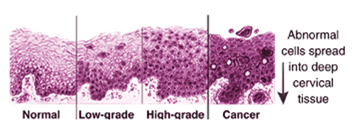
Histological Changes from Healthy to Cancer
By Anand Datta, M.D., Ph.D.
Morphological characters for interpretation — what does the screener look for?
Pap smears are subjective evaluations. Not every screener reaches the same exact conclusions. Compare the normal example to the slight changes Dr. Datta gives in his examples of ASQUS that label the latter “not normal” so that he is unable to sign the smear out as “Normal” or “Negative.”
 Reading pap smears requires experience, training, time (15 minutes per slide on average) and can be fatiguing. The law caps the number as 60 slides which can be read per 24 hours. Pathologists are required to review 10% of all read by cytotechnologist slides and ALL hi grade and cancer slides.
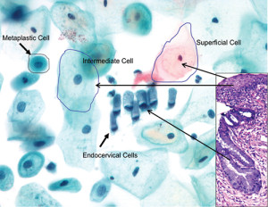
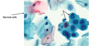
Normal Pap Smears (image to the left)
1. Superficial squamous epithelial cells: pin point nucleus with large pink and blue cells.
2. Intermediate squamous epithelial cells: slightly large nucleus in large blue cell.
3. Endocervical cells are occasionally found as part of a normal Pap smear.
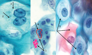
 Atypical Squamous Cells of Undetermined Significance (ASC-US)
The squamous epithelial cells are less matured, with large nucleus instead of normal pinpoint nucleus in large pink cells. Most of these cells have two abnormal nucleus in one cell. The nucleus appears rough with irregular margins.
This case is not really negative, since few cells looks to be abnormal in the midst of many normal cells.
If not monitored some cases which will progress to High-grade or cervical cancer.
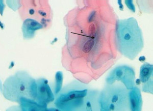
 Low Grade Squamous Intra-epithielial Lesion (LGSIL)
1# Large cells which are immature, with large nucleus, and the nucleus is at least 5 times the normal (intermediate) cell nucleus as indicated by the red dots.
2# Many cells have more than one nucleus.
Nucleus margins are usually irregular #4, and have a halo #3 around them. #5 Cells are alarming to the pathologist.
 High-Grade Squamous Intra-epithelial Lesion (HGSIL)
The HGSIL cells #1 are smaller than normal cells, have a very large nucleus, the nucleus has irregular margins, most of the nucleus is dark in color. If not actual cancer, they require treatment before progressing to cancer.
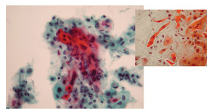 Cervical Squamous Cell Cancer (SCC)
Cervical squamous cell cancer show highly abnormal cells, with very large nucleus in a small cell, nuclear margins are irregular and stain very dark bluish green. This must be treated before it escapes the boundaries of the cervix and invades nearby organs. Some tumor cells are orange due to keratin. Keratin in cervical epithelium is abnormal.
In addition to his volunteer work for HSC, Dr. Anand Datta, Ph.D. directs a clinical lab at The National Laboratory for Clinical Research. He works closely on clinical cancer trials with NCI. Dr. Datta lives in Maryland with his wife Debbie and two young toddlers.


